
Mass Spectrometry Peptide Verification Explained

TL;DR:
- Mass spectrometry verifies peptide identity, sequence, and purity by analyzing mass-to-charge ratios and fragment patterns. It complements chromatographic methods, with MS/MS providing critical sequence confirmation, especially for modified or isobaric peptides. Proper interpretation of spectral data and detailed COAs ensure reliable peptide verification for research applications.
Mass spectrometry peptide verification is defined as the analytical process of confirming peptide identity, sequence, and purity by measuring mass-to-charge ratios (m/z) and fragment ion patterns, then matching these data against theoretical models or reference databases. This process is the standard method for unambiguous peptide identity confirmation in biochemical and molecular biology research, where chromatographic methods alone cannot distinguish co-eluting species or structurally similar sequences. Tandem mass spectrometry (MS/MS) extends this capability by fragmenting selected precursor ions and generating sequence-specific ion series, providing confirmation that intact mass measurement cannot supply. Instruments including Orbitrap analyzers, triple quadrupole systems, and time-of-flight (TOF) platforms each serve defined roles in this workflow. Certificates of Analysis (COAs) from peptide suppliers document the resulting MS data, and researchers who understand how to read these records can make procurement and experimental decisions with substantially greater confidence.
What is the typical workflow for mass spectrometry peptide verification?
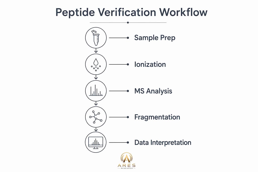
The standard workflow for peptide verification by mass spectrometry proceeds through five defined stages: sample preparation, chromatographic separation, ionization, mass analysis, and data interpretation. Each stage introduces variables that affect the quality and interpretability of the final spectral data, making procedural discipline at every step a prerequisite for reliable results.
Sample preparation and LC separation
Sample preparation begins with dissolution of the peptide in an appropriate solvent, typically aqueous acetonitrile with 0.1% formic acid, followed by desalting using C18 solid-phase extraction cartridges or ZipTip pipette tips to remove salts and matrix interferences. For complex mixtures or proteolytic digests, enzymatic digestion with trypsin, Lys-C, or Glu-C generates peptides of predictable mass and charge state distributions. Liquid chromatography (LC) separation, most commonly reversed-phase LC on C18 columns, precedes MS acquisition and resolves co-eluting species that would otherwise produce overlapping spectra. High-resolution MS combined with LC separation resolves co-eluting impurities effectively, a capability that HPLC purity measurement alone cannot replicate.

Ionization methods: ESI and MALDI
Ionization converts neutral peptide molecules into gas-phase ions detectable by the mass analyzer. ESI-MS produces multiply charged ions facilitating deconvolution to neutral mass, while MALDI-TOF generates predominantly singly charged ions and is faster for purity confirmation of peptides above approximately 2000 Da. ESI is the dominant ionization method for LC-coupled workflows because it operates continuously and interfaces directly with liquid-phase separations. MALDI is preferred for rapid screening of purified peptide fractions where throughput outweighs the need for chromatographic resolution.
MS and MS/MS instrumentation
Orbitrap-based instruments such as the Thermo Scientific Orbitrap Exploris 480 deliver mass accuracy below 5 ppm, which is the threshold at which database searches produce specific, low-false-positive identifications. Triple quadrupole instruments, including the SCIEX QTRAP series, are optimized for targeted quantification using selected reaction monitoring (SRM) and multiple reaction monitoring (MRM) modes. Ion trap and Q-TOF instruments occupy intermediate positions, offering MS/MS capability with moderate resolution. The choice of instrument determines the mass accuracy, fragmentation control, and sensitivity available for a given verification task.
Database searching and peptide-spectrum matching
Following data acquisition, raw spectra are searched against sequence databases or theoretical fragment ion lists using software platforms such as Mascot, Sequest, or MaxQuant. Fragment ion matching within strict mass tolerances of 10 ppm or less reduces false positive identifications and is the standard criterion for high-confidence peptide-spectrum matches (PSMs). Each PSM receives a score reflecting the quality of the match between observed and theoretical fragment ions, and statistical thresholds such as false discovery rate (FDR) control the proportion of incorrect identifications in the final dataset. Researchers must report these parameters explicitly when publishing or archiving verification data.
Pro Tip: When submitting peptides for verification, request that the vendor provide raw spectral files alongside the COA summary. Summary pass/fail flags do not contain the fragment ion detail needed to resolve ambiguous identifications or troubleshoot downstream assay failures.
How do fragmentation methods differ and why are they critical for peptide verification?
Fragmentation method selection determines which ion types are generated, which sequence regions are covered, and whether labile modifications survive the dissociation process. No single fragmentation technique provides complete sequence information for all peptide classes, which is why modern instruments increasingly support multiple dissociation modes within a single acquisition.
CID and HCD: the standard approaches
Collision-induced dissociation (CID) and higher-energy collisional dissociation (HCD) are the most widely deployed fragmentation methods in LC-MS/MS workflows. Both techniques accelerate precursor ions into an inert collision gas, producing predominantly b-ions (N-terminal fragments) and y-ions (C-terminal fragments) through cleavage of peptide backbone amide bonds. HCD, implemented on Orbitrap instruments, delivers higher-energy collisions and produces cleaner, more complete b/y ion series than low-energy CID in ion traps. The speed and predictability of CID and HCD make them the default choice for routine peptide sequencing and database-driven identification.
ETD and ECD: preserving labile modifications
Electron transfer dissociation (ETD) and electron capture dissociation (ECD) operate through fundamentally different mechanisms, transferring electrons to multiply charged precursor ions and generating c-ions and z-ions through N-Cα bond cleavage. ETD and ECD preserve labile post-translational modifications better than CID, improving sequence coverage and identification confidence for phosphorylated and glycosylated peptides. This preservation occurs because the radical-driven fragmentation pathway does not require vibrational energy redistribution, which is the mechanism responsible for modification loss under CID conditions. ETD requires highly charged precursors (z ≥ 3), making it most effective for longer peptides and intact proteins.
UVPD and hybrid fragmentation strategies
Ultraviolet photodissociation (UVPD), implemented on instruments such as the Thermo Scientific Orbitrap Eclipse with a 213 nm laser, generates a, b, c, x, y, and z ions simultaneously, producing the most extensive sequence coverage of any single fragmentation method. This breadth is particularly valuable for peptides containing unusual amino acids, non-natural residues, or multiple modifications that confound standard b/y ion interpretation. Complementary fragmentation methods improve identification confidence and sequence coverage beyond CID alone, and hybrid acquisition schemes that combine HCD with ETD or UVPD in a single run are now standard on high-end instruments. The practical implication is that researchers working with modified peptides should specify the fragmentation mode when requesting MS verification from suppliers or contract laboratories.
| Fragmentation method | Ion types produced | Best application | Modification preservation |
|---|---|---|---|
| CID | b, y | Routine sequencing, unmodified peptides | Low (labile groups lost) |
| HCD | b, y (high energy) | High-throughput proteomics, database searching | Low to moderate |
| ETD | c, z | Phosphopeptides, glycopeptides, large peptides | High |
| ECD | c, z | Intact protein top-down, labile PTMs | High |
| UVPD | a, b, c, x, y, z | Non-standard residues, complex modifications | High |
Pro Tip: For peptides carrying phosphorylation or O-GlcNAc glycosylation, request ETD or EThcD (electron transfer combined with HCD supplemental activation) fragmentation data specifically. Standard CID spectra from these peptides frequently show neutral loss of the modification rather than backbone fragmentation, which prevents unambiguous site localization.
What are the key interpretative considerations when verifying peptides using mass spectrometry data?
Spectral interpretation requires understanding mass accuracy, charge state assignment, isotope pattern analysis, and the specific limitations of each data type. Researchers who treat MS verification as a binary pass/fail check rather than a quantitative analytical result will miss the information needed to detect impurities, confirm modification sites, or resolve ambiguous identifications.
Mass accuracy and error tolerances
Mass spectrometry analyzes peptide mass-to-charge ratio with precision below 5 ppm error for high-resolution instruments, and this level of accuracy is what distinguishes a confirmed identity from a coincidental mass match. A 5 ppm error on a 2000 Da peptide corresponds to 0.01 Da, a tolerance narrow enough to exclude most isobaric interferences. Low-resolution instruments such as single quadrupoles operate at unit mass resolution (approximately 1 Da), which is sufficient for intact mass confirmation of short peptides but inadequate for distinguishing leucine from isoleucine or detecting small modifications such as deamidation (+0.984 Da). Researchers must confirm that the instrument type and reported mass accuracy are appropriate for the verification claim being made.
Charge states and isotope pattern interpretation
ESI generates multiply charged ions, and the charge state (z) of a detected ion must be correctly assigned to calculate the neutral monoisotopic mass. The isotope envelope, the pattern of peaks separated by 1/z Da corresponding to 13C isotope contributions, provides an independent confirmation of charge state assignment. Monoisotopic mass is the preferred reporting standard for peptides below approximately 4000 Da, while average mass is used for larger species where the monoisotopic peak is not resolved. Discrepancies between observed and expected isotope patterns indicate the presence of co-eluting species or adduct formation, both of which require investigation before accepting a verification result.
Reading COAs for MS identity data
Peptide COAs should document the following MS-specific fields for each lot:
- Observed monoisotopic or average mass with the corresponding theoretical value and mass error in Da or ppm
- Charge state(s) detected confirming that the reported m/z values are correctly assigned
- Ionization method (ESI or MALDI) and instrument platform used for the measurement
- MS/MS fragmentation data where available, including the fragmentation method and key fragment ions matched
- HPLC purity percentage as a complementary metric, with the explicit understanding that retention time and HPLC purity alone cannot confirm molecular identity
COAs typically document lot number, HPLC purity, observed vs. expected mass, charge states, and MS/MS fragmentation data, and any COA that omits the observed mass or charge state should be treated as incomplete for identity verification purposes. Aresresearchlab’s COA checklist resource provides a structured framework for evaluating these fields systematically.
Common pitfalls: isobaric peptides and co-elution
Isobaric peptides share the same nominal mass but differ in sequence or modification site, and intact mass measurement alone cannot distinguish them. For example, peptides containing leucine and isoleucine at different positions are isobaric at unit mass resolution and require MS/MS fragmentation for differentiation. Co-eluting impurities with similar retention times produce chimeric spectra in which fragment ions from multiple species are superimposed, reducing PSM scores and potentially generating false sequence assignments. Mass tolerance and ion type filters in peptide-spectrum matching are foundational parameters that must be reported for meaningful interpretation of verification results, and ignoring tight tolerance criteria risks coincidental matches and false positives.
What practical applications and best practices ensure reliable peptide verification in biochemical research?
Applying mass spectrometry verification systematically across procurement, batch acceptance, and experimental validation requires defined procedures rather than ad hoc checks. The following practices reflect the operational standards that distinguish rigorous peptide research workflows from those susceptible to identity-related experimental failures.
-
Integrate MS data with HPLC purity assessments at the point of procurement. MS is required for peptide identity verification because chromatographic methods cannot uniquely identify molecular structures due to co-eluting species with similar retention times. Researchers should require both HPLC purity data and MS identity confirmation on every COA before accepting a peptide lot for experimental use. Reviewing the HPLC purity testing guide alongside MS data provides a complete picture of compound quality.
-
Archive raw and annotated spectra linked to COA records. Raw or annotated mass spectra should be stored alongside COA summaries in laboratory records to enable future troubleshooting and reproducibility rather than relying solely on summary pass/fail indications. When a functional assay produces unexpected results, access to the original spectral data allows the researcher to determine whether the peptide identity or purity was a contributing factor.
-
Select fragmentation modes based on peptide structural properties. Unmodified peptides below 3000 Da are well-served by HCD fragmentation with Orbitrap detection. Phosphorylated, glycosylated, or otherwise modified peptides require ETD or UVPD to preserve modification sites and generate interpretable fragment ion series. Specifying the fragmentation method in purchase orders and analytical requests prevents the common situation in which a vendor provides CID data for a phosphopeptide where the phosphate group is lost before backbone fragmentation occurs.
-
Use MS data to identify peptide impurities and sequence variants. Oxidized methionine (+15.995 Da), deamidated asparagine (+0.984 Da), and truncated sequences are the most common peptide impurities detectable by high-resolution MS. These modifications alter biological activity and can confound dose-response relationships in cell-based assays. Reviewing the MS spectrum for satellite peaks at characteristic mass offsets from the main peptide ion is a standard quality control step that HPLC chromatograms cannot replace.
-
Apply MS verification data to batch-to-batch consistency monitoring. Longitudinal research programs using the same peptide across multiple lots should compare MS spectra between batches to confirm that the modification profile and impurity pattern remain consistent. Discrepancies in the fragment ion series between lots, even when HPLC purity values are equivalent, indicate synthesis or purification differences that may affect experimental reproducibility. Aresresearchlab’s peptide purity grade standards provide a reference framework for evaluating batch consistency against defined analytical criteria.
Key takeaways
Mass spectrometry peptide verification requires intact mass confirmation, MS/MS fragmentation data, and strict mass tolerance criteria to deliver unambiguous sequence identity and purity assessment.
| Point | Details |
|---|---|
| MS/MS is required for sequence confirmation | Intact mass alone cannot distinguish isobaric peptides or confirm modification site localization. |
| Fragmentation method must match peptide type | CID/HCD suits unmodified peptides; ETD or UVPD is required for labile modifications like phosphorylation. |
| COAs must include observed mass and charge state | COAs lacking these fields are incomplete for identity verification and should not be accepted. |
| Mass accuracy below 5 ppm defines high-resolution MS | Unit-mass instruments cannot resolve deamidation or distinguish leucine from isoleucine. |
| Archive raw spectra, not just pass/fail flags | Stored spectral data enables troubleshooting of downstream assay failures linked to peptide identity. |
Where mass spectrometry peptide verification is heading
From our perspective at Aresresearchlab, the most consequential development in peptide MS verification over the past two years is not a new instrument platform but the integration of deep learning models into spectral interpretation workflows. Tools such as Prosit and AlphaFold-MS-derived fragment predictors now generate theoretical MS/MS spectra with accuracy that approaches experimental measurement, enabling confident identification of peptides for which no reference spectrum exists in conventional databases. This matters practically because researchers working with non-tryptic peptides, synthetic analogs, or modified sequences have historically faced a gap between instrument capability and software interpretation. That gap is closing.
We have also observed that the field continues to underestimate the interpretive burden placed on researchers who receive only a COA summary rather than annotated spectral data. A vendor-reported “MS: Pass” flag tells the researcher that the observed mass fell within an acceptable tolerance window. It does not confirm that the correct charge state was assigned, that no co-eluting impurity contributed to the signal, or that the fragmentation pattern matches the expected sequence. Researchers who accept summary flags without requesting underlying spectral files are accepting a level of ambiguity that is inconsistent with rigorous experimental design.
The ongoing challenge of labile modifications, particularly O-GlcNAc glycosylation and sulfation, remains genuinely difficult even with ETD and UVPD. Hybrid fragmentation strategies and ion mobility separation, as implemented on instruments like the Waters SELECT SERIES Cyclic IMS, are producing meaningful improvements in these cases, but the workflows are not yet standardized across laboratories. Researchers working in this space should treat MS verification of heavily modified peptides as an iterative process rather than a single-pass confirmation.
— Ares
Verify your peptides with confidence using Aresresearchlab resources

Aresresearchlab supplies high-purity research peptides with third-party tested COAs that include observed mass, charge state data, and HPLC purity results for every lot. Researchers who need to evaluate compound quality before committing to an experimental program can consult Aresresearchlab’s compound grading standards to understand the analytical criteria applied at each purity tier. The COA interpretation checklist provides a field-by-field guide for evaluating MS identity data, HPLC purity, and lot-specific documentation against the standards described in this article. Both resources are designed for researchers who require analytical transparency rather than summary-level quality assurances.
FAQ
What does mass spectrometry measure in peptide verification?
Mass spectrometry measures the mass-to-charge ratio (m/z) of ionized peptide molecules, enabling calculation of the neutral monoisotopic mass for comparison against the theoretical value. High-resolution instruments achieve mass accuracy below 5 ppm, sufficient to confirm peptide identity and detect modifications such as oxidation or deamidation.
Why is MS/MS fragmentation necessary for peptide sequence confirmation?
Intact mass alone cannot distinguish isomers or peptides differing by labile modifications, making MS/MS fragmentation the required method for sequence-level identity confirmation. Fragment ion series (b/y from CID/HCD, c/z from ETD/ECD) map directly to amino acid positions and provide unambiguous sequence evidence.
What is the difference between ESI and MALDI ionization for peptides?
ESI produces multiply charged ions compatible with LC-coupled workflows and is the standard method for complex mixture analysis, while MALDI generates predominantly singly charged ions and is faster for single-peptide purity screening above 2000 Da. Both methods are valid provided the COA reports observed mass, charge state, and mass error.
How should researchers evaluate the MS data on a peptide COA?
Researchers should confirm that the COA reports the observed monoisotopic or average mass, the charge state(s) detected, the instrument platform, and the mass error in ppm or Da relative to the theoretical value. COAs that provide only a pass/fail flag without these fields do not supply sufficient data for independent identity verification.
Can HPLC purity replace mass spectrometry for peptide identity confirmation?
HPLC purity cannot replace mass spectrometry for identity confirmation because chromatographic retention times do not uniquely identify molecular structure. Two structurally distinct peptides can co-elute at the same retention time with identical apparent purity, making MS the required orthogonal method for unambiguous identity assignment.